MRI Protocols
Worlds Largest MRI Protocol and MRI Techniques Database
Showing posts with label
Imaging planes of the Ankle
.
Show all posts
Showing posts with label
Imaging planes of the Ankle
.
Show all posts
Imaging planes of the Ankle
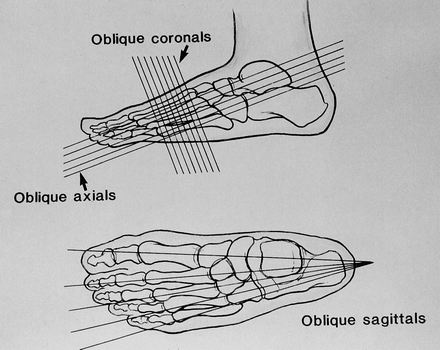
Drawings of the foot illustrate the oblique axial, oblique coronal, and oblique sagittal imaging planes.
Imaging planes of the Ankle
-->
Older Posts
Home
Subscribe to:
Posts (Atom)
Popular Posts
The Difference between CT Angiogram and MR Angiogram
Positioning of the patient in Fetal MRI
--> Patients are positioned in the supine or left decubitus position. Claustrophobic reactions have not been observed to a higher...
Thumb MRI imaging planes
Axial Plane: Prescribe plane perpendicular to midshaft of the proximal phalanx of the thumb. Scan from 1st carp-metacarpal joint th...
PECTORALIS MUSCLE MRI & ANATOMY
Normal MRI Anatomy The pectoralis tendon is best seen on axial T1 and T2-weighted images as a curvilinear low-signal band inserting o...
MR Arthrogram Protocol SHOULDER
MR Arthrogram Protocol Axial T1 SE Axial T1 SE (fat sat) Sagittal Obl T1 SE (fat sat) Coronal Obl T1 SE (fat sat) Coronal Obl T2 TSE ...
ELBOW MRI ANATOMY
Purpose of diffusion-weighted imaging (DWI) in Stoke
Early identification of ischemic stroke: diffusion restriction may be seen within minutes following the onset of ischemia Correlates wel...
Advantages and Disadvantages of fMRI
Like any technique, fMRI has advantages and disadvantages, and in order to be useful, the experiments that employ it must be carefully desi...
MRI BRAIN AXIAL ANATOMY - DETAIL
Saturation Bands in MRI Scan
Regional Saturation Bands Regional saturation employs a 90° RF pulse which, when combined with a gradient orthogonal to the imaging p...