Axial and coronal Images should include sternum and humeral head.Positioning the patient prone will reduce breathing motion artifacts.
Axial Plane: Scan from superior humeral head through the xyphoid process. The field of view should include the sternum medially, and the humeral shaft laterally.
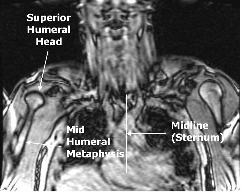

Coronal Plane: Prescribe a plane parallel to the line from sternum through anterior humerus. Scan from skin through posterior humeral head. Field of view should include the sternum and humerus.


Sagittal Plane: Prescribe plane perpendicular to coronal plane. Scan From sternum through lateral aspect of humerus.


