MRI Protocols
Worlds Largest MRI Protocol and MRI Techniques Database
Imaging planes of the Ankle
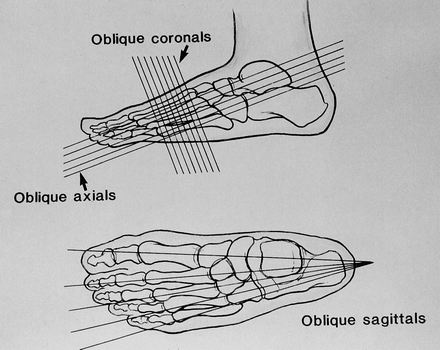
Drawings of the foot illustrate the oblique axial, oblique coronal, and oblique sagittal imaging planes.
Imaging planes of the Ankle
-->
No comments:
Post a Comment
Newer Post
Older Post
Home
View mobile version
Subscribe to:
Post Comments (Atom)
Popular Posts
MRI brachial plexus anatomy
Achilles tendon MRI images
Normal and a rupture Achilles tendon MRI images Achilles tendon MRI image-yellow arrow Green-normal Achilles tendon MRI image ...
Thumb MRI imaging planes
Axial Plane: Prescribe plane perpendicular to midshaft of the proximal phalanx of the thumb. Scan from 1st carp-metacarpal joint th...
Trigeminal Neuralgia or Facial Hemispasm MRI Protocol
Sagital T1 (whole head left to right) Axial Flair (brain) Ax T2 hi-res 3D FSE (symphysis of mandible to superior orbit) Cornonal T2...
KNEE MRI PROTOCOL & REFERENCE IMAGES
Prescribing sagittal images. Images are obtained no more than 10° oblique to a perpendicular to a line connecting the ...
CISS-MRI Sequence
High Resolution Brainstem Imaging CISS (Constructive Interference Steady State) CISS is a strongly T2 weighted GRE sequence. In essen...
Problems and pitfalls of the MRI angiogram technique
As with any imaging technique, MRA has its own artifacts and problems which must be recognized in order to avoid misdiagnosis....
THIGH (QUADRICEPS) MRI Protocol
THIGH (QUADRICEPS) - ACUTE INJURY ROUTINE PROTOCOL Check with patient re: site of tear and mark with oil capsule(s) if appropriate. Area...
MR Imaging and Outcome of TBI (Traumatic Brain Injury)
MR Imaging and Outcome of TBI MR imaging is more sensitive than CT for detecting abnormalities in the brain after TBI .This modality is...
L Spine with SI joint screening MRI Reporting Format
MRI of the LUMBAR SPINE and SI Joints. Clinical Details: Observations The lumbar vertebrae show normal marrow signal intensity. ...


No comments:
Post a Comment