- MRI is indispensable for the accurate staging of avascular necrosis (AVN), because images clearly depict the size of the lesion, and gross estimates of the stage of disease can be made (see Radiograph for the radiographic and radiologic staging systems for AVN).
- MRI allows sequential evaluation of asymptomatic lesions that are undetectable on plain radiographs.
- MRI facilitates better response to treatment because, with the use of MRI, avascular necrosis (AVN) is diagnosed at an earlier stage, and therapeutic measures are more successful the earlier they are begun.
- MRI may help guide interventional procedures such as core decompression, may demonstrate response of the femoral head to treatment, and may detect the joint effusions and bone edema that often accompany avascular necrosis (AVN)
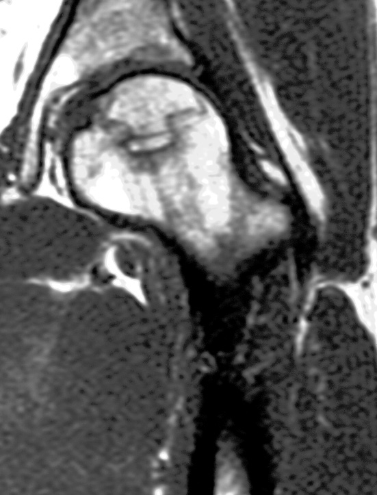
In the early stages of the disease, there may not be any alteration of the normal signal intensity of the femural head. The first sign of AVN is nonspecific: diffuse areas of decreased signal intensity are seen in the normally high-signal-intensity fatty marrow on T1-weighted images. This is thought to be due to edema within the marrow. Focal findings along the anterosuperior aspect of the femural head are more specific: low-signal-intensity bands or lines within the femoral head are seen surrounding the area that corresponds to ischemic bone on T1- and T2-weighted images. The band is thick on T1-weighted images and is thinner and accompanied by a second, innerband of high signal intensity on T2-weighted images. The appearance on T2-weighted images is known as the “double-line sign” and is considered highly specific for AVN. This band is believed to represent the reactive interface that separates normal marrow from infarcted marrow.The signal intensity of the central infarcted bone corresponds to areas of bone necrosis seen at histologic examination. High signal intensity on T1-weighted images and low signal intensity on T2-weighted images are seen within areas of necrosis when viable, fatty marrow is still present With prolonged ischemia and necrosis, the necrotic bone has a signal intensity pattern resembling that of fluid, with low signal intensity on T1-weighted images and high signal intensity on T2-weighted images. Finally, when fibrosis and sclerosis of the involved bone occurs, it is reflected by low signal intensity on both T1- and T2-weighted images. Secondary signs and sequelae of AVN can also be seen at MR imaging. Joint effusion or cartilaginous thinning may be present. Progression of AVN leads to instability of the femural head with fragmentation and eventual collapse.


No comments:
Post a Comment