MRI Protocols
Worlds Largest MRI Protocol and MRI Techniques Database
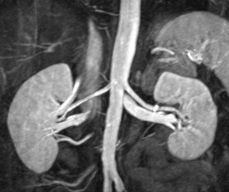
Renal MRA Protocol
Coronal SSFSE(No fat sat. 5-6mm slices)
Axial VIBE
3D Cor FLASH(Try to get effective thickness less than 1.5 mm.Want to include celiac and SMA)
20 cc Gadolinium contrast -Timing Run
3D Cor FLASH(Two measures (with 7 sec between))
Axial VIBE
Optional Cine PC
-->
No comments:
Post a Comment
Newer Post
Older Post
Home
Subscribe to:
Post Comments (Atom)
Popular Posts
The Difference between CT Angiogram and MR Angiogram
MRI brachial plexus anatomy
Positioning of the patient in Fetal MRI
--> Patients are positioned in the supine or left decubitus position. Claustrophobic reactions have not been observed to a higher...
PECTORALIS MUSCLE MRI & ANATOMY
Normal MRI Anatomy The pectoralis tendon is best seen on axial T1 and T2-weighted images as a curvilinear low-signal band inserting o...
Achilles tendon MRI images
Normal and a rupture Achilles tendon MRI images Achilles tendon MRI image-yellow arrow Green-normal Achilles tendon MRI image ...
ELBOW MRI ANATOMY
SHOULDER MRI PROTOCOL & REFERENCE LINES
--> Routine shoulder protocols are the most variable, but most include SHOULDER MRI SEQUENCES ...
OPTIC CHIASM in lateral BRAIN MRI
MRI BRAIN AXIAL ANATOMY - DETAIL
MR Arthrogram Protocol SHOULDER
MR Arthrogram Protocol Axial T1 SE Axial T1 SE (fat sat) Sagittal Obl T1 SE (fat sat) Coronal Obl T1 SE (fat sat) Coronal Obl T2 TSE ...


No comments:
Post a Comment