ANKLE AXIAL IMAGING PLANE
Axial ankle mri planning/referrence plane is parallel to axis of calcaneus.
Technical notes:
Scan ankle from distal tibia through subcutaneous soft tissues (include plantar fascia).
Cover from inferior cortex of the fifth metatarsal and cover up as far as possible ( approx. five slices above the tibiotalar joint)
Parallel Sat Bands
Cover from 3-4 Slices above the inferior margin of the tibiotalar joint (the
joint is best seen posteriorly) down as far as the slices go


ANKLE SAGITAL REFERENCE LINES-PLANNING
Prescribe plane with line parallel to talus, angled perpendicular to talar dome. Perpendicular to COR
Technical notes:
Cover ankle from medial through lateral malleolus.
Angled perpendicular to talar dome
Cover at least 1 slice out of both malleoli
All of the plantar soft tissues should be included in the FOV
Superior Sat Band


ANKLE CORONAL REFERENCE IMAGES-PLANNING
Prescribe plane perpendicular to axial imaging plane. Use axial LOC and angle perpendicular to the inner cortex of the medial malleolus
Technical notes:
- Scan ankle from calcaneus through metatarsal bases.
- Use sagittal LOC and angle parallel to distal tibial shaft and cover from the talonavicular joint to at least 2 slices posterior to the talus
- The COR PD FS sequence is thicker and can cover more of the talonavicular joint anteriorly and plantar fascia posteriorly
- All of the plantar(inferior) soft tissues should be included in the FOV
- Superior Sat Band


Ankle tendons and ligaments are best imaged with the foot plantar flexed 15-30°. So don't use the chimney in the coil. Position the calcaneus in the middle of the coil and the relaxed foot will fit comfortably in the coil at the correct degree of plantar flexion.


 T1 SAGITAL
 SAGITAL IR
Sagittal T1 and IR allow for the evaluation of the Achilles tendon, plantar aponeurosis, talar dome, subtalar facets and Sinus Tarsi.
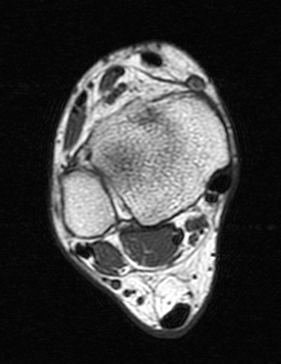
 AXIAL PD FAT SAT FSE
Axial PD fat suppression evaluates the tendons and ligaments of the ankle particularly after acute/subacute injuries. It also is sensitive to talar dome osteochondral defects. Alternatively, a T2 sequence can be used to eliminate magic angle artifact that may occur as the tendons travel around the malleolar turns
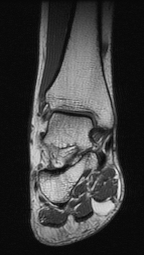
 COR OBLIQUE T1
The coronal oblique T1 sequence follows the tendons of the ankle around the malleolar turns and also evaluates the medial ankle ligaments.
 COR PD FSE FAT SAT
The coronal oblique PD fat suppressed sequence follows the tendons of the ankle around the malleolar turns and is particularly important in evaluation of the Posterior tibialis tendon.
Ankle MR Arthrogram


 Arthrogram sequences of the ankle evaluate the talar cartilage and allow for staging of ostechondral defects of the talar dome. They can also better evaluate the joint capsule and its boundaries such as the anterior talofibular ligament
Coronal T1 spin echo (SE)
 General anatomic assessment of the ankle. The tibiotalar articulation, as well as the tibiotalar ligaments are assessed. Assessment of the bone marrow on T1 is important also.
Axial T1 spin echo (SE)
Ankle tendons, muscles, general ankle anatomy all re-evaluated with this sequence. Assessment of the bone marrow on T1 is important as mentioned above.
Axial STIR
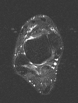
This sequence is the best to screen the bones to make sure there is not a subtle bone edema or fluid collections that is not visible on other sequences.


No comments:
Post a Comment