MRI Protocols
Worlds Largest MRI Protocol and MRI Techniques Database
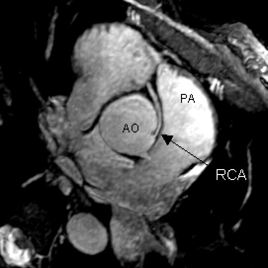
Cardiac MRI ANATOMY
-->
Cardiac MRI ANATOMY
[;
No comments:
Post a Comment
Newer Post
Older Post
Home
Subscribe to:
Post Comments (Atom)
Popular Posts
The Difference between CT Angiogram and MR Angiogram
PECTORALIS MUSCLE MRI & ANATOMY
Normal MRI Anatomy The pectoralis tendon is best seen on axial T1 and T2-weighted images as a curvilinear low-signal band inserting o...
Steady-state versus spoiled gradient echo imaging in Bright Blood Cardiac MRI Sequences
In gradient echo (GRE) imaging, the TR is often shorter than the T2 of most tissues, and the transverse magnetization will not have fully d...
ELBOW MRI REFERENCE LINES
IN THIS ARTICLE: AXIAL REFERENCE LINE CORONAL REFERENCE LINE SAGITAL REFERENCE LINE AXIAL REFERENCE LINE - Perpendicular t...
Advantages and Disadvantages of fMRI
Like any technique, fMRI has advantages and disadvantages, and in order to be useful, the experiments that employ it must be carefully desi...
Nuclear Spin and Behaviour in a Magnetic Field
Electromanetism tells us that a current carrying conductor e.g. a piece of wire, produces a magnetic field encircling it. When the wire is...
STERNO CLAVICULAR JOINTS MRI PROTOCOL
The sternum and sternoclavicular joints are difficult to evaluate with plain radiographs. The value of CT in assessing lesions of the...
ELBOW MRI ANATOMY
ABER Protocol for MRI Shoulder
ABER Protocol for MRI Shoulder Align from coronal scout perpendicular to glenohumeral joint line (perpendicular to glenoid) Ideally th...
Problems with MRI
It may not be possible, or safe, to have a MRI scan if you have any of these items: Cardiac pacemaker Surgical clips in your head (part...


 [;
[;


No comments:
Post a Comment