Plain "MRI Protocol for Meningitis"
Sag T1
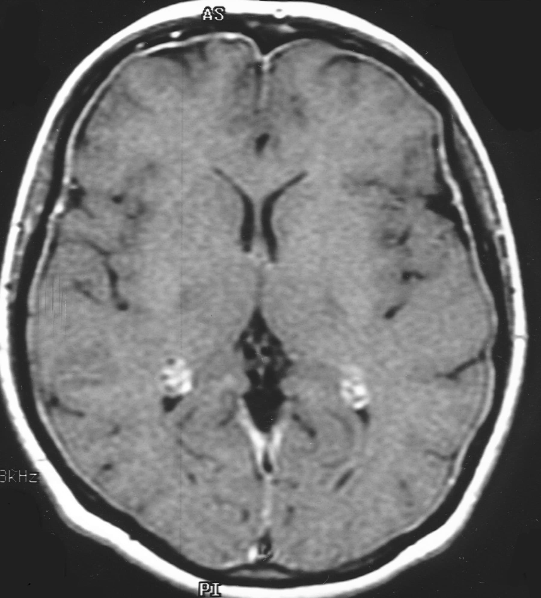
Ax T1
Ax T2 FSE/TSE
Ax FLAIR FSE/TSE
Ax DWI / ADC / B0
Cor T2 FSE/TSE
Post Contrast "MRI Protocol for Meningitis"
T1 Axial
T1 Coronal
FLAIR Axial
"MRI Protocol for Meningitis" Technical Notes
- Coronal and sagittal thin-section, heavily T2-weighted MRIs may show CSF leaks, which may be the source of infection in cases of recurrent meningitis.
- Plain and contrast-enhanced MRIs help to depict the complications of meningitis. Such complications include empyema/effusion, cerebritis/abscess, venous thrombosis, venous and arterial infarcts, ventriculitis, hydrocephalus, and edema (with or without cerebral herniation).
- The imaging features of meningitis are non-specific, demonstrating abnormal meningeal enhancement. MRI is superior to CT in the evaluation of patients with suspected meningitis
- Add FLAIR post gad in suspected meningeal disease.
- For brainstem and midline lesions get sagittal post gad instead of coronal.
- For pineal lesions add thin sagittal T2 and T1 pre and post gad images.
- Single voxel spectroscopy (TE 35 and 144) on all new mass lesions
- Multi voxel only on suspected gliomas. For follow-up use TE 144.


T1 weighted pre and post-contrast MRI's demonstrate non-specific abnormal meningeal enhancement.


No comments:
Post a Comment