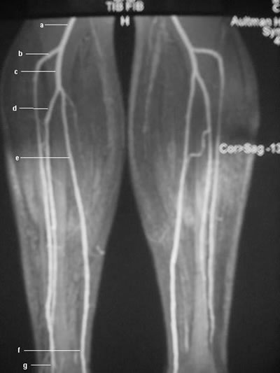
Magnetic resonance angiogram (MRA) obtained by using the bolus-chase technique shows the normal anatomy of the lower-extremity arterial vasculature, including the popliteal artery (a), the anterior tibial artery (b), the tibioperoneal trunk (c), the peroneal artery (d), and the posterior tibial artery (e).
Subscribe to:
Post Comments (Atom)
Popular Posts
-
Normal and a rupture Achilles tendon MRI images Achilles tendon MRI image-yellow arrow Green-normal Achilles tendon MRI image ...
-
Normal MRI Anatomy The pectoralis tendon is best seen on axial T1 and T2-weighted images as a curvilinear low-signal band inserting o...
-
--> This technique is entirely dependent upon the availability of a small Field of View surface coil (see below). If your unit d...
-
Sagital T1 (whole head left to right) Axial Flair (brain) Ax T2 hi-res 3D FSE (symphysis of mandible to superior orbit) Cornonal T2...
-
--> Patients are positioned in the supine or left decubitus position. Claustrophobic reactions have not been observed to a higher...
-
Prostrate MRI Appearence T1‐weighted MR images, the normal prostate gland demonstrates homogeneous intermediate to‐ low signal inten...
-
MRI SI Joint Protocol COR T1 COR STIR Axial STIR (2 STACKS) Axial/Coronal PD Fat Sat SAG T1 SAG STIR POST CONTRAST ...
-
Sagital T1 (whole head left to right) Axial Flair (brain) Ax T2 hi-res 3D FSE (symphysis of mandible to superior orbit) Cornonal T2...
No comments:
Post a Comment