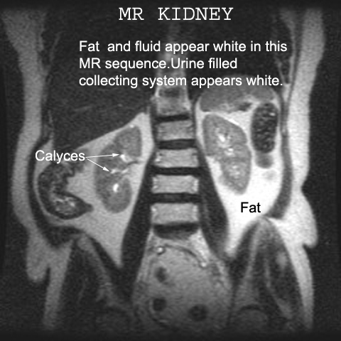
 Renal Collecting system appearence in MRI
Subscribe to:
Post Comments (Atom)
Popular Posts
-
Normal and a rupture Achilles tendon MRI images Achilles tendon MRI image-yellow arrow Green-normal Achilles tendon MRI image ...
-
--> Patients are positioned in the supine or left decubitus position. Claustrophobic reactions have not been observed to a higher...
-
Normal MRI Anatomy The pectoralis tendon is best seen on axial T1 and T2-weighted images as a curvilinear low-signal band inserting o...
-
 3-PLANE LOC AXIAL T1 AXIAL T2 FS CORONAL T1 CORONAL STIR Axial T2 high resolution FIESTA ( Facial Nerves in IAC for any space occu...
 -
Magnetic resonance imaging (MRI) cisternography depends on heavily T2-weighted sequences with fat suppression. CSF appears as a bright s...
-
MRI Prostrate Spectroscopy SINGLE VOXEL Planning --> MRI Prostrate Spectroscopy MULTI VOXEL Planning
-
 SAGITAL ORBIT MRI PLANNING REFERRAL LINES AXIAL ORBIT MRI PLANNING REFERRAL LINES CORONAL ORBIT MRI PLANNING REFERRAL ...


No comments:
Post a Comment