 |
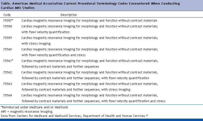
| American Medical Association current procedural terminology (CPT) for CARDIAC MRI |
Popular Posts
-
The sternum and sternoclavicular joints are difficult to evaluate with plain radiographs. The value of CT in assessing lesions of the...
-
 ABER Protocol for MRI Shoulder Align from coronal scout perpendicular to glenohumeral joint line (perpendicular to glenoid) Ideally th...
 -
 3-PLANE LOC AXIAL T1 AXIAL T2 FS CORONAL T1 CORONAL STIR Axial T2 high resolution FIESTA ( Facial Nerves in IAC for any space occu...
 -
 Prescribing sagittal images. Images are obtained no more than 10° oblique to a perpendicular to a line connecting the ...
 -
--> Patients are positioned in the supine or left decubitus position. Claustrophobic reactions have not been observed to a higher...
-
 --> Routine shoulder protocols are the most variable, but most include SHOULDER MRI SEQUENCES ...
 -
Normal MRI Anatomy The pectoralis tendon is best seen on axial T1 and T2-weighted images as a curvilinear low-signal band inserting o...
-
Early identification of ischemic stroke: diffusion restriction may be seen within minutes following the onset of ischemia Correlates wel...
-
Magnetic resonance imaging (MRI) cisternography depends on heavily T2-weighted sequences with fat suppression. CSF appears as a bright s...


No comments:
Post a Comment