T2-weighted images are used for both detection and characterization of focal liver lesions. A routine liver imaging protocol includes a Fast Spin-Echo (FSE) T2-weighted sequence. FSE sequences use long repetition times (TRs), greater than 2000 msec. Selection of echo time (TE) is important since image contrast is dependent on this value. The optimal TE used clinically varies from 80-100 msec. Even longer TE's are used for characterization of
hemangiomas and cysts. The addition of fat suppression to a T2- weighted FSE sequence further improves image quality by reducing motion artifact and improving tissue contrast.
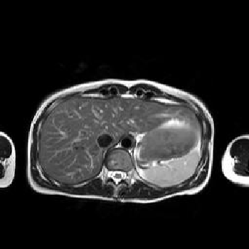
T2-weighted Images
• Used for both lesion detection and characterization.
• SE or FSE acceptable.
• Fat saturation reduces motion artifact and improves tissue contrast.
• Respiratory triggering or compensation is useful.
• Optimal TE varies from 80-105 msec.
T2-weighted SE images have been traditionally used for liver MR and they have been shown to have utility for both lesion detection and characterization. Unfortunately, since only a single echo is obtained during a long TR, data acquisition times for a single T2-weighted SE study commonly exceed 13-18 minutes. Because of the long acquisition times, SE images are
sensitive to motion-related artifacts. Motion compensation methods such as respiratory triggering are helpful but often further increase scan times. T2-weighted FSE imaging generates T2-weighted images more rapidly than SE imaging. Following each 90 degree excitation pulse, FSE imaging uses 180 degree refocusing pulses to generate multiple echoes, instead of acquiring a single echo as is done in SE imaging. This markedly reduces acquisition
times from 13-18 min down to 3-6 minutes. However, since multiple echoes cannot be
obtained simultaneously, they are clustered together around a desired “effective TE“
(TEef). This causes FSE T2-weighted images to have somewhat different image contrast
than SE images. Fat is usually bright on FSE images while it is intermediate to dark on SE images. Depending on the number of echoes obtained (echo train length), FSE images may have some blurring; limiting the echo train length to 4-8 will minimize this while still


No comments:
Post a Comment