IN THIS ARTICLE:
- MRI Brain Axial Planning/Reference Lines
- MRI Brain Coronal Planning/Reference Lines
- MRI Brain Sagital Planning/Reference Lines
MRI Brain Axial Planning/Reference Lines


LOOK AT TO THE RED AC AND YELLOW PC ..
- Axial MRI Brain slices are positioned parallel to the bicommissural line,which links to the anterior and posterior commisure(yellow line).
- Axial Brain mri slices can be also position parallel to a line linking the floor of the sella turcica to the fastigium of the fourth ventricle .
- Another Axial Brain MRI refference line is,position the slices parallel to a line linking the inferior borders of the genu and splenium of the corpus callosum
These imaging planes differ by a few degress. It is important that if you are a Technologist/Radiologist set a standard imaging plane from these 3 suggestions and therefore you can compare the follow-up scans compare to the baseline study and even you can compare any of the scans performed under you.
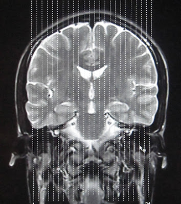
MRI Brain Coronal Planning/Reference Lines
 |
| MRI BRAIN CORONAL REFFERENCE LINE |
For CORONAL MRI BRAIN an imaging plane parallel to the brainstem is preferred in sagital blocaliser, in axial localizer the mid scan line is made parallel to the the line joining the right and left internal auditory meatus or posterior aspect of orbits. This gives symmetrical coronal images. Make sure that the scan lines cover the whole brain parenchyma from frontal lobe to the posterior aspect of cerebellum.
MRI Brain Sagital Planning/Reference Lines

Use the coronal scout to plan the true midsagittal image parallel to the falx and other midline structures.
On a true midsagittal image a line is drawn through the hypophysis and the roof of the fourth ventricle (fastigium).
This is called the HYFA: hypophysis-fastigium line.
This is called the HYFA: hypophysis-fastigium line.
The HYFA line should pass through the interhemispheric fissure in axial localizer and in coronal localizer, the scan lines are made parrallel to the interhemispheric fissure so that the sagittal images cover whole brain parenchyma from right sylvian fissure to the left sylvian fissure.


READ MORE ABOUT:


No comments:
Post a Comment