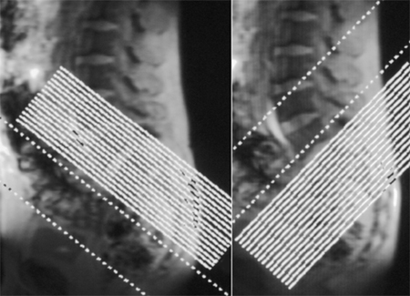
The image shows the orientation of scan planes on an initial lateral scout view of the region. To the left semi-coronal and to the right semi-axial slice orientation.
|
|


Popular Posts
-
 Prescribing sagittal images. Images are obtained no more than 10° oblique to a perpendicular to a line connecting the ...
 -
Normal MRI Anatomy The pectoralis tendon is best seen on axial T1 and T2-weighted images as a curvilinear low-signal band inserting o...
-
 3-PLANE LOC AXIAL T1 AXIAL T2 FS CORONAL T1 CORONAL STIR Axial T2 high resolution FIESTA ( Facial Nerves in IAC for any space occu...
 -
 ABER Protocol for MRI Shoulder Align from coronal scout perpendicular to glenohumeral joint line (perpendicular to glenoid) Ideally th...
 -
 --> Routine shoulder protocols are the most variable, but most include SHOULDER MRI SEQUENCES ...
 -
Early identification of ischemic stroke: diffusion restriction may be seen within minutes following the onset of ischemia Correlates wel...
-
--> Patients are positioned in the supine or left decubitus position. Claustrophobic reactions have not been observed to a higher...
-
Magnetic resonance imaging (MRI) cisternography depends on heavily T2-weighted sequences with fat suppression. CSF appears as a bright s...
-
Axial Plane: Prescribe plane perpendicular to midshaft of the proximal phalanx of the thumb. Scan from 1st carp-metacarpal joint th...
-
Position patient prone, head first into the scanner with arms by the side. Head to one side and chest relaxed onto the breast coil. ...


No comments:
Post a Comment