-->
Plain "Cerebral Abscess MRI Protocol"
MR spectroscopy : elevation of a succinate peak is relatively specific but not present in all abscesses ; high lactate, acetate, alanine, valine, leucine, and isoleucine levels peak may be present ; Cho / Crn and NAA peaks are reduced.Magnetic resonance (MR) spectroscopy may be helpful in the differential diagnosis of toxoplasmosis versus CNS lymphoma. CNS lymphoma generally shows a mild pattern of elevated lipid and lactate peaks, with a prominent choline peak with some other normal metabolites. In toxoplasmosis, there are elevated lipid and lactate peaks, while other normal brain metabolites are nearly absent.
Plain "Cerebral Abscess MRI Protocol"
- T1-Sagittal head
- T1 SE-Axial head
- FLAIR Axial head
- T2 FSE Axial head
- DWI/ADC
Post Contrast "Cerebral Abscess MRI Protocol"
- T1 SE-Axial post-gadolinium (This is essentially the same sequence as the pre-gadolinium scan, but can be performed with the addition of a flow compensation (FC) pulse to better delineate the cerebral vessels, and a magnetization transfer (MT) pulse to optimize enhancing lesion detection.)
- T1-coronal post-gadolinium head
- MR perfusion
- MR spectroscopy
Radiology notes:"Cerebral Abscess MRI Protocol"
MR perfusion : rCBV is reduced in the surrounding oedema c.f. to both normal white matter and tumour oedema seen in high grade gliomas .MR spectroscopy : elevation of a succinate peak is relatively specific but not present in all abscesses ; high lactate, acetate, alanine, valine, leucine, and isoleucine levels peak may be present ; Cho / Crn and NAA peaks are reduced.Magnetic resonance (MR) spectroscopy may be helpful in the differential diagnosis of toxoplasmosis versus CNS lymphoma. CNS lymphoma generally shows a mild pattern of elevated lipid and lactate peaks, with a prominent choline peak with some other normal metabolites. In toxoplasmosis, there are elevated lipid and lactate peaks, while other normal brain metabolites are nearly absent.
Diffusion-weighted MRI may be useful in differentiating abscess MRI features from necrotic tumor.
Early cerebritis: swollen, edematous, areas of necrosis, ill-define margins; nonspecific (tumor, infarct)
Late cerebritis: increased central necrosis, thick irregular contrast enhancement, high on FLAIR, T2, and DWI
Early capsule: within 2 weeks, walled off capsule, necrotic center, enhancing rim
Late capsule: more defined rim, multiloculated, capsule is low on T2, markedly high on DWI

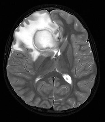


_--_showing_a_small_ring-enhancing_lesion_with_mild_surrounding_edema_adjacent_to_the_ventricular_catheter_and_ventricular_dilatation..jpg)

Brain Abscess MRI findings Classification
- Early cerebritis stage-The early cerebritis stage presents as an ill-defined subcortical hyperintense zone that can be noted on T2-weighted imaging. Contrast-enhanced T1-weighted studies demonstrate poorly delineated enhancing areas within the isointense to mildly hypointense edematous region.
- Late cerebritis stage-During the late cerebritis stage, the central necrotic area is hyperintense to brain tissue on proton-density and T2-weighted sequences. Peripheral edema is common. The rim enhances intensely following contrast administration. Satellite lesions may be demonstrated.
- Early and late capsule stages-During the early and late capsule stages, the collagenous abscess capsule is visible prior to contrast as a comparatively thin-walled, isointense to slightly hyperintense ring that becomes hypointense on T2-weighted MRIs.Diffusion-weighted imaging aids in depiction of specific features of a brain abscess. If a cerebral abscess ruptures into the ventricular system, diffusion-weighted images demonstrate specific patterns.Purulent material within the ventricle appears similar to that of the central abscess cavity, with a strongly hyperintense signal on diffusion-weighted images.
MRI Features in different Stages


1 comment:
There are a lot of misconceptions about what it means to be living with Herpes simplex virus (HSV). Ultimately, everyone’s lives are different – how you cope with your diagnosis and how you move forward is up to you.
The 21st of June 2015 was the day I was diagnosed with Herpes simplex virus (HSV). l was so worried that I cried out endlessly. On the way there I was crying and shaking the steering wheel, I just didn’t know what I should do.
After that day, my depression was at all-time high, but slowly, I started to do my own research, looking at what I should do next and then last month, i came across a website which was about cure for Herpes simplex virus (HSV) and other STD's.
I took the contact details from the website and contact the doctor (Dr. Francisco. B.), and after much inquiry, we came to a conclusion and she sent a herbal medicine to me through the courier delivery service, and she also gave me guidelines on how to take the medicine.
After 9 days of taking the medicine, i discovered that all painful sores in the genital area, the itching, painful urination and tender lumps in the groin was no more, and i went for a medical test, and i was so amazed to see my test result showing NEGATIVE. I then went to another hospital, and my test result is still showing NEGATIVE till date.
I think life has given me another chance to be happy and free, I now do what I can to inspire others through these battles. For anyone out there affected with such disease please contact Dr. Francisco .B. with the below link;
Email: franciscobrain8@gmail.com
Post a Comment