ANKLE MRI Protocol


Without contrast "ANKLE MRI"  Sequences  
  • Sagital  T1
  • Sagital  T2 FSE STIR (or fat sat)
  • Axial  T2 FSE STIR (or fat sat)
  • Coronal  T2 STIR (or fat sat)
  • Coronal  T1/PD FSE
  • Axial  PD FSE  Fat Sat

Post Contrast "ANKLE MRI" Sequences 
  • Axial  FMPSPGR  Fat Sat  PRE/POST
  • Sagital T1 SE NON  Fat Sat
  • Axial  T1 SE  Fat Sat
  • Coronal   T1 SE  Fat Sat

"ANKLE MRI" ARTHROGRAM PROTOCOL  
  • Sagital  T1 SE Fat Sat
  • Sagital STIR
  • Axial   T1 SE  Fat Sat
  • Axial PD FSE NON  Fat Sat
  • Coronal   T1 SE  Fat Sat
  • Coronal T2 FSE  Fat Sat

MR images of the ISCHEMIC STROKE

MR IMAGING IN ISCHEMIC STROKE CASE (DIFFUSION AND PERFUSION).

Foot MRI Protocol


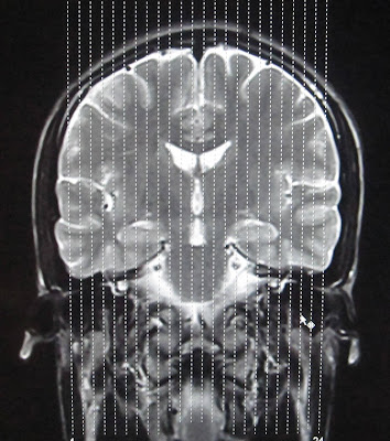
Without contrast FOOT MRI Sequences
  •  AX (LONG AXIS) T1
  •  AX (LONG AXIS) STIR
  •  COR (SHORT AXIS) STIR
  •  COR (SHORT AXIS) T2
  •  SAG STIR
  •  SAG T1
  •  THIN SLICE AXIAL PD FAT SAT

POST CONTRAST FOOT MRI
GAD- COR FMPSPGR FAT SAT PRE/POST GAD

POSITIONING of the Patient for FOOT MRI
  • Prone and foot is plantar flexed (Preferred Method)
  • Supine and toes sticking out of coil (Alternative Method)


TECHNICAL NOTES
The foot and ankle are among the hardest of all areas to image because of the complex three-dimensional anatomy. Magnetic resonance imaging (MRI), with its multiplanar capabilities, excellent soft-tissue contrast, ability to image bone marrow, noninvasiveness, and lack of ionizing radiation, has become a valuable tool in evaluating patients with foot and ankle problems. MRI is more specific than bone scintigraphy and provides more information than ultrasound and computed tomography. Arthroscopy of the ankle is limited to the articular surface and joint space. MRI allows a global evaluation of the bones, tendons, ligaments, and other structures with a single examination that exceeds the capabilities of all other available techniques. This monograph was written to provide a useful guide to basic technique, indications, positioning, anatomy, and interpretation of foot and ankle MRI. The first part describes the performance of the MRI examination with reference to the positioning of the foot, types of coils, and advantages and disadvantages of the different sequences and imaging planes. The next section was written by an experienced foot and ankle orthopedic surgeon and outlines the indications for MRI for the common foot and ankle symptom complexes and the information that the surgeon hopes to obtain from the study. This is followed by a review of pertinent anatomy, as it applies to imaging, with emphasis on osseous structures, ligaments, tendons, and muscles. The final section is a comprehensive review of the common pathologic conditions encountered in the foot and ankle. We hope that radiologists and radiologists-in-training find this article a useful reference tool and gain a better understanding of this complex area of musculoskeletal imaging.


READ MORE:


Foot MRI Reference Lines

CERVIX MRI Protocol

MRI CERVIX without Contrast


  • Axial T1 SE
  • Axial oblique  T2 FSE Fat Sat
  • Sagital T1 SE
  • Sagital T2 FSE

Post contrast MRI CERVIX

  • Dynamic contrast-enhanced T1-weighted images (small field of view) in the sagittal/axial oblique-single pre contrast run with 4 post contrast acquisitions.
  • 3D T1 Fat Sat Sagital/Axial oblique(Optional)

Note:Dynamic contrast-enhanced MRI improves detection of small tumors and helps in differentiating tumor recurrence from radiation fibrosis. The use of contrast medium is not necessary for cervical cancer examinations because it does not improve staging accuracy compared with unenhanced T2-weighted images 

Thoracic spine MRI Protocol


T-spine mri protocol without contrast

  • T2 Sagittal FSE
  • T1 Sagittal SE
  • T2 Sagitta STIR
  • T2 Axial FSE
  • T1 Axial SE

POST CONTRAST T-SPINE MRI

  • Post-Gd sagittal T1 SE with fat saturation
  • Post-Gd axial T1 SE with fat saturation

Optional MRI T-SPINE SEQUENCES

  • Coronal T2 FSE (scoliosis)


MRI Brain Reference Lines/Images


IN THIS ARTICLE:
  • MRI Brain Axial Planning/Reference Lines
  • MRI Brain Coronal Planning/Reference Lines
  • MRI Brain Sagital Planning/Reference Lines


MRI Brain Axial Planning/Reference Lines

LOOK AT TO THE RED AC AND YELLOW PC ..

  • Axial MRI Brain slices are positioned parallel to the bicommissural line,which links to the anterior and posterior commisure(yellow line). 
  • Axial Brain mri slices can be also position parallel to a line linking the floor of the sella turcica to the fastigium of the fourth ventricle .
  • Another Axial Brain MRI refference line is,position the slices parallel to a line linking the inferior borders of the genu and splenium of the corpus callosum
These imaging planes differ by a few degress. It is important that if you are a Technologist/Radiologist set a standard imaging plane from these 3 suggestions and therefore you can compare the follow-up scans compare to the baseline study and even you can compare any of the scans performed under you.

MRI Brain Coronal Planning/Reference Lines
MRI BRAIN CORONAL REFFERENCE LINE
For CORONAL MRI BRAIN an imaging plane parallel to the brainstem is preferred in sagital blocaliser, in axial localizer the mid scan line is made parallel to the the line joining the right and left internal auditory meatus or posterior aspect of orbits. This gives symmetrical coronal images. Make sure that the scan lines cover the whole brain parenchyma from frontal lobe to the posterior aspect of cerebellum. 

MRI Brain Sagital Planning/Reference Lines
Use the coronal scout to plan the true midsagittal image parallel to the falx and other midline structures.
On a true midsagittal image a line is drawn through the hypophysis and the roof of the fourth ventricle (fastigium).
This is called the HYFA: hypophysis-fastigium line.
 
The HYFA line should pass through the interhemispheric fissure in axial localizer and in coronal localizer, the scan lines are made parrallel to the interhemispheric fissure so that the sagittal images cover whole brain parenchyma from  right sylvian fissure to the left sylvian fissure.


READ MORE ABOUT:


Pituitary MRI Protocol


IMAGING PLANES OF CARDIAC MRI

Horizontal long axis: known as a 4-chamber view.
Short axis plane-multiple/contiguous
Vertical long axis: known as a 2-chamber view


Short axis plane-Short Axis Stack (SAS)


Vertical Long Axis (VLA)


Left Ventricular Outflow Tract


Right Ventricular Outflow Tract


Pulmonary Artery


Phase Contrast Aorta


Phase Contrast PA


IMAGING PLANES OF CARDIAC MRI
sagittal single-shot fast spin-echo image is used as an initial localizer for coronal imaging
Coronal single-shot fast spin echo-aortic valve
Four-chamber gradient-echo image oblique axial plane serves as a localizer for the short-axis view
Coronal single-shot fast spin-echo image obtained off the sagittal plane. To localize the atrioventricular valve level, an imaging plane is prescribed along a line connecting the cardiac apex and the level of the middle of the atrioventricular valve, as shown.
Four-chamber gradient-echo image-cross sectional depiction of the atrioventricular valves
The 3-chamber view can be prescribed from the left ventricular outflow tract view of a short-axis view. This view displays the aortic and mitral valves immediately adjacent to one another.
Left ventricular outflow view: Steady-state gradient echo (SSFP [steady-state free precession]) left ventricular outflow view demonstrates a bicuspid aortic valve and an aortic jet secondary to aortic stenosis.


MR Enterography Protocol

Magnetic resonance (MR) enterography has the potential to safely and noninvasively meet the imaging needs of patients with bowel diseases without exposing them to ionizing radiation. MR Enterography can provide high resolution dynamic images of bowel motility without exposing the patient to radiation.


Patient preparation
 Bowel prep day before – low residue diet, fluids, laxative. 
Overnight fasting or NPO 4-6 hrs prior to study
Technical preparations

All adults and kids should get 3 bottles of oral contrast (VoLumen (low-conc barium (0.1% weight/volume) that contains sorbitol) - each bottle is 450 ml. 
The patient receives an IV antispasmodic to stop bowel motion. 

Prone position preferred to spread and better visualize the bowel loops.
Spasmolytics are useful for reducing bowel peristalsis and motion artifacts. Reduction of peristalsis is most important for fast gradient-echo sequences performed after the administration of intravenous contrast material and also may help reduce intraluminal flow artifacts on half-Fourier acquisition single-shot turbo spin-echo images.
MR Enterography Sequences

  • Scano  
  • Coronal T2 SSFSE  Abdomen&Pelvis
  • Axial T2 SSFSE  Abdomen&Pelvis
  • Coronal 2D FIESTA Abdomen&Pelvis
  • Axial2D FIESTA Fat Sat Abdomen&Pelvis
  • Axial LAVA BH  Abdomen&Pelvis


POST CONTRAST
  • COR LAVA BH MP  Abdomen&Pelvis
  • Axial LAVA BH  Abdomen&Pelvis(90 seconds after injection and covering the entire abdomen)
  • Coronal 3D-MRCP upper and lower (done for bowel, not for bile ducts)


Optional:eDWI coronal with 1400 b-value,demonstrating Crohn’s disease,Lymphadenopathy

ADC mapping will be useful to assess ADC marking Malpighi cell proliferation, thus we can identify the surge of infl ammatory Crohn’s disease.Also LAVA images fused with ADC maps is possible.


SSFSE-Single shot fast spin echo, FIESTA-fast imaging employing steady state precession,LAVA-Liver Acquisition with Volume Acquisition(3 dimensional spoiled gradient echo pulse sequence)


Pectoralis Muscle MRI Image planes


Axial and coronal Images should include sternum and humeral head.Positioning the patient prone will reduce breathing motion artifacts.


Axial Plane: Scan from superior humeral head through the xyphoid process.  The field of view should include the sternum medially, and the humeral shaft laterally.


Coronal Plane: Prescribe a plane parallel to the line from sternum through anterior humerus. Scan from skin through posterior humeral head.  Field of view should include the sternum and humerus.

Sagittal Plane: Prescribe plane perpendicular to coronal plane. Scan From sternum through lateral aspect of humerus.


MRI Orbit Protocol


  • Coronal STIR Orbits
  • Coronal T2/T1 Fat Sat Orbits
  • Axial T2 Fat Sat Orbits
  • Axial T1 Orbits
  • Sagital T2 Fat Sat orbits(Parallel to optic nerve)

POST CONTRAST "MRI ORBIT PROTOCOL"
  • POST AX T1 Brain
  • POST COR T1 Fat Sat
  • POST AX T1 Orbits

OPTIONAL IF "BRAIN &ORBIT MRI" DONE IN SAME STUDY INCLUDE THE FOLLOWING SEQUENCES
  • Sagital T1 BRAIN
  • Axial FLAIR
  • Axial DWI
  • (If separate ADC map:) Axial  ADC
  • AX T2 Brain

Note: Images through orbits include from globe through optic chiasm

SI Joint MRI Protocol


MRI SI Joint Protocol
  • COR T1
  • COR STIR
  • Axial STIR (2 STACKS)
  • Axial/Coronal PD Fat Sat
  • SAG T1
  • SAG STIR
POST CONTRAST  

  • Post gad Axial T1 Fat Sat
  • Post gad Oblique Coronal T1 Fat Sat

CORONAL IMAGING PLANE OF SI JOINTS MRI

Oblique Coronal is angled parallel to the sacrum.FOV for the coronal images should include the entire sacrum and coccyx, and both SI joints

3D MRCP Pancreas Technique


SOURCE: http://imageradiology.blogspot.com/2012/06/3d-mrcp-pancreas.html
The 3D TSE sequence can produce high-spatial-resolution MRCP images . Thin sections without a slice gap allow better assessment of small stones, side branches of the main pancreatic duct, and intrahepatic bile ducts. Three-dimensional TSE MRCP may be performed as a series of breath-holds or during free breathing. We acquire 1–2 mm, contiguous slices during free breathing and use the navigator-echo technique to reduce motion effects. The main disadvantage of this technique is the relatively long acquisition time. In addition, navigator-based triggering requires uniform and regular breathing cycles for optimal image quality. If the patient has rapid or irregular breathing, the image quality may be impaired. An alternative method of producing 3D MRCP images is to use a TSE sequence with a 90° flip-back pulse. This sequence is called FRFSE (fast recovery fast spin-echo), DRIVE, or RESTORE. The unique feature of this sequence is that after a long echo-train, the residual transverse magnetization is refocused into a final spin-echo and then flipped along the z-axis by a –90° fast recovery pulse . This accelerates relaxation of the longitudinal magnetization, leading to a reduction in TR without a loss of SNR. It is possible to perform breath-hold 3D MRCP with this sequence. However, the number of slices that may be obtained is substantially less than with respiratory-triggered versions of 3D MRCP.
-->
Three-dimensional volumetric MRCP images are of superior quality and give better delineation of pancreaticobiliary anatomy than conventional 2D images and have the added advantage of multiplanar and postprocessing capabilities.

READ MORE:
Dynamic Coronal MRCP
Secretin-Enhanced MRCP Protocol


Secretin-Enhanced MRCP Protocol


--> Secretin is a naturally occurring 27-amino acid polypeptide released by ductal mucosa in response to acid in the lumen. Secretin increases bicarbonate and pancreatic fluid secretion by the exocrine cells. Secretin relaxes the sphincter of Oddi and opens pancreatic duct orifices.
Secretin is injected intravenously at the time of the MRCP. Images are then taken every 30 seconds for 10 minutes. Maximum output of pancreatic fluid is optimal between 6 to 8 minutes. A dynamic image and video is created from a 3-D rendering by the radiologist. This image sequence examines the pancreas response to stimulation. The excess pancreatic fluid/bicarbonate will resonate a sharper image of the pancreas. Static MRCP’s turn into dynamic images with pancreatic stimulation.
Secretin enhanced MRCP (S-MRCP) relies on dynamic responses of the main pancreatic duct to secrete fluid after Secretin stimulation, which causes the improvement of visual clarity in sharpening pancreatic imaging Secretin is a polypeptide hormone secreted by duodenal mucosa in response to luminal acid . It induces pancreatic secretion of water and bicarbonate. In the first 5–7 minutes, the tone of the sphincter of Oddi is increased. These effects result in temporary distention of the pancreatic ducts. Synthetic human secretin  is given IV over 1 minute to avoid potential abdominal pain that may occur with a bolus injection. An adult dose of 16 μg (0.2 μg/kg body weight in children) is used. At the commencement of injection, a baseline scan is obtained, followed by coronal SSFSE images (2-second scanning time) every 30 seconds for 10 minutes. In healthy subjects, the maximal effect of IV secretin is between 7–10 minutes 

READ MORE:
Dynamic Coronal MRCP
3D MRCP Pancreas Technique

Diffusion tensor imaging in spinal cord Injury


Source:  http://imageradiology.blogspot.com/2011/11/diffusion-tensor-imaging-in-spinal-cord.html 
Spinal cord injuries result in damage to the myelinated fibers of the spinal cord and/or nerve roots, causing myelopathy. 
 There are various causes of spinal cord injuries, e.g., trauma, tumor, and demyelination. These injuries can cause damage to the central gray matter, involving interneurons and motor neurons. Pathologically, such spinal cord insult can cause Wallerian degenerationeither above or below the level of injury. MRI can detect these changes as increased signal intensity on T2W(T2 weighted) images. However diffusion tensor imaging (DTI) has the potential to detect abnormalities in the spinal cord, even in cases where routine MRI (Magnetic resonance imaging) may be normal.


DTI is being widely used in the brain for various applications. DTI in diffuse axonal injury has been extensively studied.  Recently, the feasibility of tensor imaging in the spinal cord has been tested both in the cervical and the lower cords. The clinical application of tensor imaging in spinal cord lesions due to trauma, tumors, and inflammation has shown the usefulness of this technique. DTI has even been able to demonstrate displaced white matter tracts or their involvement by lesions in the cord, thus helping treatment planning and follow-up of cases. 
The greatest advantage of tensor imaging is that it can show changes in white matter tracts even in cases where routine imaging is normal. In diffuse axonal injury, where routine CT (Computed tomography) scan and MRI were normal, there was reduction in diffusion anisotropy after 24 h, suggesting axonal injury.  Similarly, in demyelinating disease such as multiple sclerosis, reduced FA in the cervical cord has been demonstrated in patients as compared to controls, although routine MRI imaging was normal. Also, it has been well documented that signal changes seen on routine MRI may not correlate with neurological deficits and clinical findings, whereas DTI has been shown to correlate with motor deficits. 
Wallerian degeneration above or below the injury level has also been demonstrated on pathological examination.  Buss et al, has shown that there is sequential loss of myelin proteins during Wallerian degeneration after spinal cord injury that can be seen years after injury. Tensor imaging spinal cord contusion has shown evolving changes in the ADC with recovery in ADC values with time suggesting that recovery from spinal cord injury is a dynamic process that goes on for years. 
Tensor imaging has the potential to noninvasively identify axonal regeneration after stem cell therapy.
In conclusion, DTI in the spinal cord is a feasible technique. It can detect Wallerian degeneration, which is not detected on routine imaging.It correlates well with motor deficits and is a predictor of long-term motor recovery.


READ MORE CLICK THE FOLLOWING LINKS:


HIP Joint MR-Arthrogram Protocol


HIP MR-ARTHROGRAM
For dedicated imaging of a single hip during arthrogram, a smaller field of view with a dedicated coil or flexible surface coil is used, allowing for visualization of smaller structures such as the labrum.  Notice the decreased SNR due to the smaller field of view.


Sequence for HIP Arthrogram


  • Coronal - both hips T1
  • Axial - affected hip      T1 Fat Sat
  • Axial -affected hip       PD Fat Sat
  • Coronal -affected hip   T1 Fat Sat
  • Coronal -affected hip PD Fat Sat COR
  • Sagittal -affected hip T1 Fat Sat SAG
  • Sagittal -affected hip PD Fat Sat SAG
  • Optional 
  • Oblique Axial T2 parallel to femoral neck


Read more regaring HIP Joint:


HIP Joint Anatomy

Hip Joint MRI Reference Lines


Axial Imaging Plane


  • Prescribe plane parallel line bisecting lesser trochanters and/or acetabular roofs. Scan from iliac crests through lesser trochanter.
  • Use COR T1 and angle parallel to femoral heads/acetabuli
  • Cover from 2-4 slices above acetabuli down close to lesser trochanters
  • Parallel Sat Bands
Coronal Imaging Plane


FOR HIGH RESOLUTION WITH FLEX COIL PLANNING


  • Prescribe plane parallel femoral heads.
  • Scan from ischium through pubicsymphyses
  • Use Axial LOC and angle parallel through femoral heads
  • Cover from back of ischial tuberosities to at least 2 slices anterior toacetabuli (preferably to cover pubic symphysis)
  • Superior Sat bands for STIR and T1
Sagittal Imaging Plane


  • Prescribe plane perpendicular to coronal plane.Scan from acetabulum through greater trochanter.
  • Perpendicular to COR PD
  • Use COR PD and cover from outer cortex of the greater trochanter to the 
  • inner portion of the acetabulum
  • Center at Femoral Head/Neck Junction
Axial Oblique Plane


Prescribe plane parallel to femoralneck.  Scan through entire femoral neck.

  •  Use COR PD and angle parallel to femoral neck (use image with the longest medial/inferior femoral neck cortex).  This angle is usually slightly more than you think (see image).
  • Cover from 1 slice out of acetabulum superiorly to 1 slice out of 
  • acetabulum inferiorly
  • Center at Femoral Head/Neck Junction Superior Sat Ban


                                                             AXIAL OBLIQUE IMAGE-PD FSE


Read more regaring HIP Joint:


HIP Joint Anatomy


Related Posts Plugin for WordPress, Blogger...

Popular Posts