
Axial Imaging Plane


- Prescribe plane parallel line bisecting lesser trochanters and/or acetabular roofs. Scan from iliac crests through lesser trochanter.
- Use COR T1 and angle parallel to femoral heads/acetabuli
- Cover from 2-4 slices above acetabuli down close to lesser trochanters
- Parallel Sat Bands


- Prescribe plane parallel femoral heads.
- Scan from ischium through pubicsymphyses
- Use Axial LOC and angle parallel through femoral heads
- Cover from back of ischial tuberosities to at least 2 slices anterior toacetabuli (preferably to cover pubic symphysis)
- Superior Sat bands for STIR and T1
Sagittal Imaging Plane


- Prescribe plane perpendicular to coronal plane.Scan from acetabulum through greater trochanter.
- Perpendicular to COR PD
- Use COR PD and cover from outer cortex of the greater trochanter to the
- inner portion of the acetabulum
- Center at Femoral Head/Neck Junction
Axial Oblique Plane

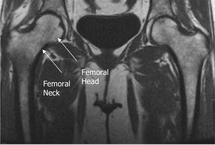

Prescribe plane parallel to femoralneck. Scan through entire femoral neck.

- Use COR PD and angle parallel to femoral neck (use image with the longest medial/inferior femoral neck cortex). This angle is usually slightly more than you think (see image).
- Cover from 1 slice out of acetabulum superiorly to 1 slice out of
- acetabulum inferiorly
- Center at Femoral Head/Neck Junction Superior Sat Ban


