Showing posts with label Pelvis-Hip Region MRI Protocols. Show all posts

Urinary Bladder MRI Protocol

  • T2 TSE Sag 4mm
  • T2 TSE Ax 4mm
  • T1 in Ax Coverage, bifurcation to pubic symphysis.
  • HASTE Cor Thru kidneys. Use body coil if necessary.
  • Fast multiplanar spoiled gradient-echo images with fat suppression Axial plain and contraast enhanced Dynamic 2mm thickness
Technical notes for "Urinary Bladder MRI Protocol"

Sagittal and coronal gadolinium-enhanced images were added if the tumor was located in the base or the dome of the bladder. 
On T2-weighted images, the normal bladder wall was identified as a hypointense line outlining the bladder lumen 
MRI with dynamic contrast administration has been shown to be superior to CT, particularly in detecting superficial and multiple tumors and in detecting extravesical tumor extension and surrounding organ invasion
 Ideally, the urinary bladder should be moderately distended during imaging. The low distension may cause misevaluation of the bladder wall thickening, and overdistension may obscure small tumors. 
-->

PELVIS VENOGRAM MRI Protocol

  • AXIAL2D Time of Flight (SUPERIOR SAT BAND)
  • AXIAL Phase Contrast
  • AXIAL  2D FIESTA  
  • Contrast enhanced MRV
 

-->


Perianal fistula MRI Protocol

NORMAL MRI ANO-RECTAL FISTULA MRI PROTOCOL
-->

  • T2 FSE 3 Planes-Axial-Coronal 5mm with intervel .5mm
  • T2 FSE Fat Sat Axial-Coronal-Sagital 5mm with intervel .5mm
  • T1 FSE Fat Sat Axial 5mm with intervel .5mm

  • POST CONTRAST 
    • T1 FSE Fat Sat Axial 5mm with intervel .5mm
    OPTIONAL SEQUENCES
    • STIR Axial/Coronal
    • 3D Dynamic Post contrast
    • T2-weighted SE with saline instillation (MR fistulography)
    TECHNICAL NOTES:
    •  Mucin-containing fistulas were recognized as tubular structures with a hyperintense signal surrounded by a hypointense rim on T2-weighted two-dimensional turbo SE images. Non–mucin-containing fistulas were recognized as tubular structures with a hypointense signal on T2-weighted images. Fluid-filled cavities were hyperintense on T2-weighted images and surrounded by a border of hypointense signal.
    • Dynamic gadolinium-enhanced imaging has been described as superior to STIR for detecting active sepsis, with the main advantage of being faster but with the additional cost of gadolinium use. MR fistulography with instillation of saline can facilitate the detection of fistula tracks, but the technique is cumbersome and depends on the existence of an external opening
    • MR imaging with an endoanal coil can also generate images with a high spatial resolution because of the very high signal-to-noise ratio near the coil. The main drawback of the endoanal MR imaging technique is that it fails to show many secondary extensions that lie beyond the range of the and this problem can be overcome by combining the endoanal coil with a phased-array coil. 
    •  Spasmolytics such as 20 mg of hyoscine butylbromide (Buscopan; Boehringer Ingelheim, Ingelheim, Germany) or 1 mg of glucagon administered intramuscularly may help to reduce motion-induced artifacts. (Hyoscine butylbromide is not licensed for use in the United States.)


    Perianal fistula imaging planes

    MRI Perianal fistula AXIAL imaging planes
    Figure 11:
    TECHNICAL NOTE: AXIAL MRI Perianal fistula is planning truly transverse according to the anal canal


    MRI Perianal fistula CORONAL imaging planes


    TECHNICAL NOTE: AXIAL MRI Perianal fistula is planning parallel to the anal canal or perpendicular to 
    the axial imaging planes.

    Anal/Rectal Cancer MRI Protocol


    • Axial-Coronal-Sagital 3plane T2 Fat Sat (some radiologists preferred T2 non fat sat-because the differentiation with the T2-weighted sequences is based on the contrast between the high-signal-intensity mesorectal fatty tissue and the rather low signal intensity of the tumor, spectral fat suppression techniques are not needed)
    • Axial T1 FSE with short TE(5mm slice thickness) / T1-weighted 3D gradient-echo sequence-Lymph node evaluation
    Post contrast MRI Anal cancer
    • T1 fat saturated Axial-Coronal-Sagital 3plane
    Technical notes:
    • 3 mm thickness slices preffered.
    • Ensure imaging planes are perfectly perpendicular the mural wall for best assessment of the wall.
    • There is not much role in the staging anal cancer and gadolinium-so for staging purpose we are not using the gad
    Angulation 
    Axial images have to be abgulated perpendicular to the axis of the tumor to avoid volume averaging.
    At first the axial images were not properly angulated. This resulted in the false impression, that the circumferential resection margin was involved on the anterior side (red circle). After proper angulation it was noted, that the CRM was not involved (yellow circle).
    Coronal sequences we can plan perpendicular to the axial planiing.

    MR Urography Protocol


    The most common MR urographic techniques for displaying the urinary tract can be divided into two categories: static-fluid MR urography and excretory MR urography.
    In static fluid MR urography the urography is done  by SSFSE heavy T2  Coronal and in excretery mr urography it is done by IV fluid excretion.

    Without Contrast Sequences
    • Localizer -Abdomen & Pelvis
    • Coronal SSFSE -Abdomen & Pelvis
    • Axial GREDIENT T1 -Abdomen & Pelvis
    • Axial T2 Fat Sat-Respiratory Triggered-Abdomen & Pelvis
    • Axial 3D GRADIENT Fat Sat
    POST DIURETIC-PRE Contrast sequences
    Now we have to admin Diuretic and acquire the sequence
    •  Ureters and Bladder Thick slab MR Urographic coronal 
    Post contrast seuences
    • 3D Fat Sat GRADIENT Dynamic Axial (pre contrast-during the time of contrast-20 sec and 45 sec acquisitions)
    • Pelvis 3D Fat Sat GRADIENT Axial(must be perform after the dynamic-ie,before the IV contrast reaches the bladder)
    • 3D Fat Sat GRADIENT Axial-Abdomen & Pelvis (Excretory phase)
    • 3D Fat Sat GRADIENT Coronal-abdomen & Pelvis (Excretory phase)

    Patient Preparation

    • Let the patient void prior to the scan
    • If possible give 200 ml normal saline IV at the start of imaging
    • If possible give oral negative contrast agents to reduce the bowel contents interpretation in the imaging.

    READ MORE ABOUT:


    CERVIX MRI Protocol

    MRI CERVIX without Contrast


    • Axial T1 SE
    • Axial oblique  T2 FSE Fat Sat
    • Sagital T1 SE
    • Sagital T2 FSE

    Post contrast MRI CERVIX

    • Dynamic contrast-enhanced T1-weighted images (small field of view) in the sagittal/axial oblique-single pre contrast run with 4 post contrast acquisitions.
    • 3D T1 Fat Sat Sagital/Axial oblique(Optional)

    Note:Dynamic contrast-enhanced MRI improves detection of small tumors and helps in differentiating tumor recurrence from radiation fibrosis. The use of contrast medium is not necessary for cervical cancer examinations because it does not improve staging accuracy compared with unenhanced T2-weighted images 

    HIP Joint MR-Arthrogram Protocol


    HIP MR-ARTHROGRAM
    For dedicated imaging of a single hip during arthrogram, a smaller field of view with a dedicated coil or flexible surface coil is used, allowing for visualization of smaller structures such as the labrum.  Notice the decreased SNR due to the smaller field of view.


    Sequence for HIP Arthrogram


    • Coronal - both hips T1
    • Axial - affected hip      T1 Fat Sat
    • Axial -affected hip       PD Fat Sat
    • Coronal -affected hip   T1 Fat Sat
    • Coronal -affected hip PD Fat Sat COR
    • Sagittal -affected hip T1 Fat Sat SAG
    • Sagittal -affected hip PD Fat Sat SAG
    • Optional 
    • Oblique Axial T2 parallel to femoral neck


    Read more regaring HIP Joint:


    HIP Joint Anatomy

    Hip Joint MRI Reference Lines


    Axial Imaging Plane


    • Prescribe plane parallel line bisecting lesser trochanters and/or acetabular roofs. Scan from iliac crests through lesser trochanter.
    • Use COR T1 and angle parallel to femoral heads/acetabuli
    • Cover from 2-4 slices above acetabuli down close to lesser trochanters
    • Parallel Sat Bands
    Coronal Imaging Plane


    FOR HIGH RESOLUTION WITH FLEX COIL PLANNING


    • Prescribe plane parallel femoral heads.
    • Scan from ischium through pubicsymphyses
    • Use Axial LOC and angle parallel through femoral heads
    • Cover from back of ischial tuberosities to at least 2 slices anterior toacetabuli (preferably to cover pubic symphysis)
    • Superior Sat bands for STIR and T1
    Sagittal Imaging Plane


    • Prescribe plane perpendicular to coronal plane.Scan from acetabulum through greater trochanter.
    • Perpendicular to COR PD
    • Use COR PD and cover from outer cortex of the greater trochanter to the 
    • inner portion of the acetabulum
    • Center at Femoral Head/Neck Junction
    Axial Oblique Plane


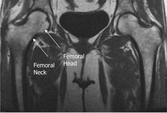
    Prescribe plane parallel to femoralneck.  Scan through entire femoral neck.

    •  Use COR PD and angle parallel to femoral neck (use image with the longest medial/inferior femoral neck cortex).  This angle is usually slightly more than you think (see image).
    • Cover from 1 slice out of acetabulum superiorly to 1 slice out of 
    • acetabulum inferiorly
    • Center at Femoral Head/Neck Junction Superior Sat Ban


                                                                 AXIAL OBLIQUE IMAGE-PD FSE


    Read more regaring HIP Joint:


    HIP Joint Anatomy


    Hip Joint MRI Protocol


    Magnetic resonance imaging (MRI) of hip joint - is one of the most promising and rapidly improving techniques of modern diagnostics. In this case, the doctor is able not only to investigate the structural and pathological changes, but also to assess the physico-chemical, pathophysiological processes of the hip joint as a whole or its individual structures.


    MRI HIP SEQUENCES

    • COR T1 SE NON FAT SAT
    • COR FSE STIR
    • AXIAL T2 FSE FAT SAT
    • AXIAL OBLIQUE PD FSE FAT SAT
    • Axial STIR
    • COR T2 FSE FAT SAT
    • SAG PD FAT SAT

    Pelvic MRI protocol : Endometriosis


    Axial T1

    Axial T2
    Fat Sat T1 Axial, Coronal & Sagittal
    This protocol can be applied if the patient is having known endometriosis.
    If suspecting any other pathological conditions please include GADOLINIUM administered T1 

    Note:IV buscopan is administered to reduce bowel peristalsis and hence improve image quality.

    Gadolinium is not particularly useful in evaluation of endometriomas  and does not improve detection of very small implants as small vessels enhance and may be misinterpreted as a small endometriotic deposit . Additionally, enhancement of endometrial deposits is variable post contrast and does not differentiate from other benign or malignant processes.

    Rectus Femoris MRI Protocol


    Protocol-Rectus Femoris
    • Axial fat suppressed T2-weighted image
    • Fat suppressed T2-weighted images parasagittal
    • Coronal STIR 
    • Sagittal fat suppressed proton density 
    • Axial and coronal T1 weighted
    • Axial fat suppressed T2-weighted
    Optional sequences
    • coronal turbo spin-echo fat-suppressed fluid-weighted images
    • sagittal fluid-weighted images 
    NOTE: The slice thickness was 4–5 mm with a 10% gap.


    Rectus femoris injuries are extremely common in athletes, particularly in soccer players, rugby player, and sprinters. Magnetic resonance imaging (MRI) plays a key role in diagnosis, prognosis, and rehabilitation of these injuries. The current article discusses current concepts in the diagnosis and treatment of rectus femoris injuries in elite athletes, including a discussion of the less well known myofascial injuries and key prognostic factors as seen at MR imaging.
    Axial and coronal MR images are optimal for visualizing the direct and indirect heads, the conjoined tendon, and the deep musculotendinous junction of the proximal rectus femoris. Tears of the deep musculotendinous junction are longitudinal, involving a long segment of the muscle. MRI features include a “bull's-eye” sign, longitudinal scar, retraction, pseudocyst, and hematoma.


    Previous SectionNext Section

    MRI Prostrate Protocol


    MRI Prostate without contrast Sequences
    -->

    • Axial T1‐weighted pelvis
    • Axial T2‐weighted pelvis
    • Sagital  T2‐weighted pelvis
    • Coronal Oblique T2‐weighted prostate
    • MR Spectroscopy of selected volumes of the prostate
    • Diffusion‐weighted imaging with ADC

    Post Contrast "MRI PROSTATE PROTOCOL"

    • Dynamic contrast enhancement Coronal Oblique small FOV

    Radiology Notes for "MRI PROSTATE PROTOCOL"
    • MRI is performed at least 3 to 4 weeks after prostate biopsy because the presence of blood products may obscure or be mistaken for prostate cancer. 
    • Prior to MRI, an intramuscular spasmolytic drug is injected to diminish bowel peristalsis. 
    • We are using combination of an endorectal and pelvic phased-array coils. The endorectal coil is inserted and inflated with approximately 60 to 80 mL of liquid per fluorocarbon.
    • Proton MRSI( Spectroscopy )reveals specific metabolic information about the prostate.
    • Dynamic contrast-enhanced MRI used to find out data reflecting tissue perfusion, micro-vessel permeability, and the extracellular leakage space can be obtained. As with other organs abnormal areas demonstrate different enhancement patterns than that of normal tissue. Typically this data is acquired with a high temporal resolution (3 to 5 seconds). This requires the trade off in the resolution of the imaging to obtain the fastest possible acquisition.
    Patient Preparation of the "MRI PROSTATE PROTOCOL"
    • NPO 4 hours prior to the examination - this reduces bowel motility.
    • Empty bladder, have patient void prior to positioning for exam. This further reduces motion and significant displacement of the organ during data acquisition. 
    • Empty rectum/sigmoid - bowel cleansing treatment or enema just prior to the MR exam may be considered to eliminate trapped air pockets that will compromise susceptibility sensitive imaging techniques (DWI, spectroscopy). 
    • Valium (5-10mg po) or Xanax (1-2 mg po) if patient is claustrophobic
    Patient positioning: Supine, feet first. The arms are positioned comfortably next to abdomen. Patient must have comfortable pillow for head and a cushion under the knees to relieve back pressure.


    T2 AXIAL

    T2 CORONAL

    T2 SAGITAL

                    DWC                                                   ADC


    MRI Prostrate Spectroscopy


     3T dual coil DCE axial
                      MRI Prostrate Dyanmic Contast Enhancement


    MRI Uterus Protocol

    SEQUENCES REQUIRED
    • Large Field-of-View Coronal SSFSE
    • Sagittal T2
    • Axial T1
    • Axial T2 (Axial to Uterus)
    • Coronal T2 (Coronal to Uterus)
    POST CONTRAST
    • Fat Sat T1 Axial
    • Fat Sat T1 Coronal
    • Fat Sat T1 Sagital
    OPTIONAL SEQUENCES
    • Axial T1 Fat Sat entire pelvis-if endometriosis suspected
    • Post contrast DYNAMIC UTERUS if UAE(uterine artery embolism)


    TECHNICAL NOTES:
    Imaging technique and patient preparation are important to obtain optimal results. Patients are usually instructed to fast for 4–6 hours before the MRI examination to limit artifact due to small-bowel peristalsis. An anti-peristaltic agent (hyoscine butyl bromide or glucagons) may be administered to the patient before imaging as an alternative to fasting. Ideally, the patient is asked to empty the bladder before going on the MR scanner. A full bladder may degrade T2-weighted images because of ghosting and motion artifacts. Patients are imaged in the supine position using a pelvic surface array multichannel coil.
    The basic MRI protocol  includes axial T1-weighted spin-echo images with a large field of view to evaluate the entire pelvis and upper abdomen for lymphadenopathy and bone marrow changes; high-resolution T2-weighted fast spin-echo (FSE) images in the axial and sagittal planes for the evaluation of the primary tumor; and dynamic contrast-enhanced T1-weighted images (small field of view) in the sagittal and axial oblique planes to evaluate the extent of myometrial and cervical involvement.


    Sagittal T2(Normal pelvis Sagital)


    • Anterior saturation band over subcutaneous fat helps to eliminate respiratory motion artifact
    • Include entire uterus and lower lumbar spine and sacrum
    • If there is pelvic pain than it may be useful to also acquire proton density (TE = 17) images at the same time to evaluate any intervertebral disc disease.

    Axial T1(Normal pelvis Axial)

    Useful to evaluate the uterine contour, lymph nodes, and bone marrow,fatty masses, hemorrogic collection/cysts, adenopahthy and muscles.
    • Cover entire pelvis
    • Place anterior saturation band
    • If endometriosis is suspected, repeat with fat saturation.

    Axial T2 (Axial to Uterus)

    This sequence shows the zonal anatomy ofthe uterus, evaluates any endometrial abnormalities and shows the relationship of any fibroids to the endometrium and identifies submucusal fibroids which may cause irregular bleeding. If adnexal mass suspected use pelvis axial instead of uterine axials.


    Coronal T2 (Coronal to Uterus)


    This sequence evaluates abnormalities of the uterus.  It is acquired coronal to the uterine fundus. Do not use fat saturation, bright fat helps to identify the outer contour of the fundus. Remember for the entire pelvis MRI my preference goes to T2 Fat Sat.


    Contrast enhancement is used to document the extent of endometrial carcinoma invasion or to detect the presence of necrosis in uterine leiomyomas. Dynamic contrast injection can be used in women who are considering uterine artery embolization (UAE) in order to evaluate the uterine arteries and the potential collateral gonadal arterial supply. 1 In the evaluation of congenital uterine anomalies, sagittal T2W images should be acquired first to determine the long-axis orientation of the uterus. Subsequently, images should then be obtained parallel to the long axis of the uterus in order to show the outer uterine contour

    MRI Protocol for Imaging of Endometrial Cancer


    MRI Protocol Endometrium

    • Axial 
      T1-weighted (upper abdomen and pelvis) 
    • Axial 
      oblique (short axis)  
      T2- FSE slice thickness < 4 mm
    • Sagittal T2-weighted FSE
    • T1-weighted 3D gradient-echo with fat saturation
    POST CONTRAST MRI ENDOMETRIUM

    • Dynamic Contrast Endometrium T1  Sagital/Oblique axial
    • T1-weighted contrast-enhanced 3D gradient-echo with fat saturation

    Note:Optimal timing to detect myometrial invasion is ~ 2 minutes following contrast injection.


    DYNAMIC PELVIS MRI & CYSTOCOLPOPROCTOGRAPHY


    Protocol

    Axial, coronal and sagittal T2 whole pelvis
    Sagittal midline T2 -3 images
    At rest
    Squeezing/Clenched
    Straining/Bearing down
    Sagittal T2 Bearing down 15 images
    Sagittal T2 single slice 1/second during evacuation
    Sagittal T2 post evacuation Valsalva

    Imaging Protocol
    Imaging with the patient in the supine position has been shown to be perfectly satisfactory for evaluating symptomatic pelvic floor weakness, despite the fact that defects are most easily identified when patients are upright . The MR imaging protocol requires no oral or intravenous contrast agents, and the examination can be completed in 15 minutes. Once familiar with signs of pelvic descent, the radiologist can complete the interpretation in 5–10 minutes.
    After the patient has voided, she is positioned on the MR imager table with a multicoil array wrapped low around the pelvis. Scout images are obtained to identify a midline sagittal section that shows the pubic symphysis, urethra, vagina, rectum, and coccyx. Next, 10-mm-thick sagittal images of the midline are obtained with a rapid half-Fourier T2-weighted imaging sequence, such as single-shot fast spin echo (SSFSE; GE Medical Systems, Milwaukee, Wis) or half-acquisition single-shot turbo spin echo (HASTE; Siemens, Iselin, NJ), and a 30-cm field of view. These images are obtained while the patient is at rest and during the Valsalva maneuver. Many patients require coaching to achieve maximal pelvic strain. Next, 5-mm-thick axial T2-weighted images of the pelvic floor centered on the puborectal muscle are obtained with a 20-cm field of view and a standard high-resolution sequence such as fast spin echo. Coronal images are optional. 
    Imaging Technique:
    In MRI of the pelvis, adequate patient preparation and a good technique with fast acquisition time are required to achieve maximum patient comfort and hence better patient compliance. The patient will be asked to void partially before the dynamic examination in order to prevent a distended urinary bladder from obscuring the pelvic structures and masking pelvic organ prolapse. Maintaining a small amount of urine in the urinary bladder improves visualization of the bladder and anterior vaginal wall prolapse. The examination is performed with a torso phased-array coil wrapped around the pelvis. Although use of an endovaginal coil may improve the spatial resolution of the fine supporting ligaments in the pelvis, it is invasive and may diminish patient acceptance and compliance [9]. The use of an endovaginal coil may distort the pelvic tissues in patients with a small pelvis. The field of view is small and often inadequate for visualization of the puborectalis. To improve visualization of the vagina and rectum, a small volume of intraluminal ultrasonic gel that has a hyperintense T2 signal may be instilled. Via a small-caliber catheter-tip syringe, 20 mL of gel may be instilled into the vagina and approximately 60–120 mL into the rectum. Although the dynamic MRI examination may be performed without endoluminal gel, doing so results in suboptimal straining that masks the degree of pelvic organ prolapse and results in inconspicuity of visceral descent.

    Ultrafast, large-field-of-view, T2-weighted sequences such as single-shot fast spin-echo (SSFSE, GE Healthcare scanners) or half-Fourier acquisition turbo spin-echo (HASTE, Siemens Medical Solutions scanners) are frequently described in dynamic MRI of the pelvic floor and are performed at our institution. Alternatively, true fast imaging in steady-state precession may be performed. The patient should be given instructions as to the proper performance of straining before the examination. Specifically, she should be told to keep her sacrum on the table and strain using only the internal organs. The images are acquired in the sagittal plane and can be viewed in a cine loop to visualize the pelvic floor and the degree of prolapse of the pelvic organs.

    For patients with a rectocele, these images should be repeated after the patient evacuates the rectal contents. The evacuation sequence can be obtained with the patient in the magnet and recorded if the magnet has been adequately prepared and the patient is able to cooperate with instructions. However, if the patient cannot evacuate in the magnet, evacuation in the commode followed by repeated imaging may be necessary. Residual contrast material will define a significant rectocele. In patients with pelvic organ prolapse, static images may be acquired in the coronal plane. These images show ballooning of the iliococcygeus muscle that often occurs with chronic constipation and perineal hernias.

    After the dynamic examination is completed, small-field-of-view (20–24 cm) T2-weighted axial fast spin-echo (FSE, GE Healthcare scanners) or axial turbo spin-echo (TSE, Siemens Medical Solutions scanners) sequences are acquired to obtain high-resolution images of the muscles and fascia of the pelvic floor and the fascial condensations supporting the urethra. Although this set of images requires approximately 4 minutes to acquire, images of the lower pelvis are resistant to breathing motion artifacts. These high-resolution axial images of the pelvis are useful in showing the relationship between the pelvic side wall and the urethra and vagina. Fat saturation is generally not applied to these sequences because the hyperintense signal of fat in the pelvis provides good contrast to the hypointense signal of the adjacent muscles, fascia, and pubic bones. The entire examination is typically completed in 20 minutes.

    Imaging Technique:

    Pelvic coil and fast T2-weighted (T2W) sequences, such as single-shot fast spin echo, half Fourier 
    acquisition turbo spin echo, or steady-state free precession sequences (true FISP) are typically used for dynamic pelvic floor MR imaging. On the T2W images, the fluid in the bowel and urine in the bladder, as well as pelvic fat have bright signal, which allows clear delineation of the pelvic organs. To achieve good visualization of the vagina and rectum, intraluminal gel (sterile lubricating gel), which gives high T2 signal, should be instilled. About 120 cc of warmed gel is instilled into the rectum, and 20 cc into the vagina. MR imaging of the pelvic floor can be performed without endoluminal contrast, however lack of vaginal and rectal distention may lead to suboptimal evaluation . Vaginal gel allows better visualization of the anterior and posterior vaginal walls, therefore improves detection of the apex of the vagina and its inversion, if present, which can be difficult to reveal otherwise. Without rectal distension with gel, adequate straining, evacuation, and rectal emptying cannot be properly documented and recto-vaginal septum defects and posterior rectal wall laxity and intrarectal intussusception may not be visualized during the exam while contributing to defecatory dysfunction in the real life. Also, non-emptying of a large rectocele may obscure an enterocele. Over distention of the bladder can mask prolapse in other compartments, therefore patients are asked to void prior to the study.Imaging is usually performed in three planes (axial, sagittal, and coronal). Images acquired in the sagittal plane at midline can be viewed in a cine loop to visualize pelvic organs descent during strain maneuvers and Valsalva. Sagittal images are used to evaluate and measure pelvic organ position at rest and their descent during strain. Coronal images are used to assess the symmetry of the levator ani muscles. Axial images are important in the assessment of the levator hiatus, the shape of the vagina, and lateral defects. Patients are imaged in the supine position or in the left lateral decubitus position in the standard closed magnet configuration. For the patients to be imaged in the upright position, the open magnet system can be used. Patients are instructed to perform different maneuvers during the scanning, such as Kegel squeeze or strain and defecation, to allow the dynamic evaluation of the pelvic floor function. The combination of gravity and rectal evacuation maximizes the stress on the pelvic floor. Rectal evacuation during MR scan in supine position is especially important due to diminished effects of gravity in this body orientation. Majority of patients can defecate in the supine position. Some patients need to have their knees bent over the pillow to 
    facilitate rectal emptying. Typically, the study is completed in less than 20 minutes.


    Dynamic pelvic MR imaging technique

    Dynamic pelvic MR imaging may be performed in either closed or open-configuration MR systems and allows for the evaluation of the pelvic floor in different positions.Al though open- conf igur a t ion MR  systems in  sitting position enable a more physiological approach to defecation, the use of such systems is limited by the lack of their world wide availability. Since Bertschinger et al (10) have shown that no clinical significant findings were missed when comparing dynamic pelvic MR in supine position with dynamic pelvic MR in sitting position, MR in supine position may be considered a reliable imaging modality for imaging the pelvic floor. In our experience, performing the examination using state-of-the-art technique, which means MR imaging at rest, at maximal contraction of the anal sphincter ( squeezing) , at  straining,as well as imaging during evacuation is probably more important than the patient position. In particular, MR imaging during defecation is of paramount importance since relevant findings may be missed when dynamic pelvic MR encompasses imaging at rest, at squeeze and at straining only . Although there is general agreement that no premedication nor oral or intrarectal preparation for bowel cleansing is necessary when dynamic pelvic MR imaging is performed, there is considerable variation in the literature with regard to the optimal examination protocol. For evaluation of the posterior compartment (i.e. the anorectum) all authorities agree that the rectum should be filled with contrast agent. The viscosity of the enema should be somehow similar to that of a normal rectum content because the manifestations of outlet obstruction may vary with fecal consistency . Hence, when performing dynamic pelvic MR, most authors use ultrasound gel for rectal enema . Other authors have proposed the use of mashed-potatoes spiked with a small amount of gadolinium-chelate for filling the rectum .The increasing use of dynamic MR imaging has shown that patients with abnormalities of the posterior pelvic compartment often reveal concomitant disorders involving the anterior as well as the middle com-partment . Therefore some authors have proposed also to tag  the vagina (i.e. the middle compartment) and/or the bladder (i.e. the anterior pelvic compartment) in order to clearly delineate the   anterior   and   the   middle   pelvic   compart - ments . In our experience, there is no need for contrast agent administration neither in the vagina nor in the bladder since MR imaging allows easily the identification of all the anatomical landmarks of the three pelvic compartments.
     Drawing of the sagittal midline view of the female pelvis shows bony landmarks and the puborectal muscle, also called the levator sling. The pubococcygeal, H, and M lines and the levator plate are delineated in red.


    Advanced Imaging Techniques

    The MR imaging protocol described above is all that is required for preoperative evaluation of the symptomatic patient with pelvic floor weakness. Several authors have described the use of upright imaging in an open-configuration MR imaging unit . Although this technique does maximize the evidence of all weakness, it rarely helps identify new defects or changes the therapeutic approach. Three-dimensional imaging has also been explored as a tool for determining levator ani volume in an attempt to select patients for either conservative therapy or surgery . At present, it remains a research technique.


    Dynamic pelvic MR imaging in sitting position


    Dynamic MR imaging of the pelvic floor in sitting  position is performed in a vertically open-configuration MR s y s t e m   s u c h   a s   t h e   0 . 5   T   s u p e r c o n d u c t i n g   o p e n -
    configuration MR system Signa SP (GE Medical Systems, Milwaukee, Wis). A wooden chair, which fits into the open space between the two magnet rings, allows imaging in the sitting position . Before the subject is placed on the seat, the rectum is filled with 300 mL of a contrast agent solution which consists of a suspending agent (mashed potatoes) spiked with 1.5 mL of an extracellular 0.5 M gadolinium-chelate contrast agent . A flexible transmit/receive radiofrequency coil is wrapped around the pelvis. On the basis of axial localizing images, a 15-mm thick multiphase T1-weighted spoiled gradient-recalled echo (GRE) sequence which transverses the rectal canal in the midsagittal plane is planned. An image update is provided every 2 seconds. Images are obtained at rest, at maximal sphincter contraction, during straining, and during defecation in the midsagittal plane. The images are analysed on a workstation in cine loop presentation. Additional gradient-echo (GRE) images are used in axial and coronal planes when a lateral rectocele or an internal rectal prolapse is suspected. Dynamic pelvic MR imaging  in supine position Dynamic pelvic MR imaging may also be performed in  supine position using nearly all commercially available closed- or open-configuration MR systems with horizontal access. When dynamic pelvic imaging is performed in these MR systems the patient is placed in supine position and a pelvic phased-array coil is used for signal transmission and/ or reception. For dynamic MR imaging in the different positions various MR sequences can be used with similar results. The basic requirement for the sequence is the necessity for a fast imaging update. Some authors have used T2-weighted single-shot fast spin-echo sequences (SSFSE) in the midsagittal plane obtained at rest, at squeezing, at straining and during defecation . Al t e r n a t i v e l y,   s t e a d y   s t a t e   f r e e   p r e c e s s i o n   ( S S F P )sequences may be used for this purpose. Our current protocol includes (SSFP) sequences obtained at rest, at squeezing, straining, and after evacuation. For assessment of the defecation we prefer the use of a T1-weighted multiphase GRE sequence since this sequence offers an imaging window long enough for continuous imaging of t h e   e v a c u a t i o n ,   e v e n   i n   p a t i e n t s  wi t h   a   p r o l o n g e d evacuation time. The choice of the enema depends on the MR sequence which is used for dynamic imaging of the pelvic floor. For SSFSE and SSFP sequences ultrasound gel is most suitable (10,13, 22, 23). If the dynamic sequences are performed with some sort of T1 weightening the rectum
    is filled with an enema which consists of ultrasound gel or mashed potatoes mixed with a small amount of gadoliniumchelate as described above. MR findings in patients with outlet obstruction Rectocele An anterior rectocele is the most frequent anatomical abnormality in patients with pelvic floor disorders and is defined as a rectal wall protrusion or bulging during defecation. The anterior wall is most commonly involved but a rectocele may also be located in the posterior rectal wall. Posterior rectoceles are also referred  as a posterior perineal hernia by some authors based on the fact that the bulging is through a puborectalis muscle defect . Two pathogenetic mechanisms are involved in the formation of rectoceles: (a) the weakness of the rectovaginal septum either congenital or after an obstetric trauma, and (b) chronic straining at defecation in constipated patients. Rectoceles may occur in up to 20% of asymptomatic women . However, there is general agreement that only rectoceles with a sagittal diameter of more than 2 cm may result in outlet obstruction, and/or the need of digital manoeuvres to empty the rectum . A clinical significant rectocele should be considered based  on the following criteria: patient history, size exceeding 2 cm in sagittal diameter, retention of contrast medium, reproducibility of the patient’s symptoms, and the need for evacuation assistance . Most of the rectoceles are diagnosed during physical examination but a reliable classification with regard to the size, emptying, and associated abnormalities are only provided by imaging examinations. Dynamic MR enables an accurate assessment of the size, location and degree of emptying of a rectocele. Using dynamic pelvic MR imaging, an anterior rectocele may be classified with regard to its size, expressed as the depth of wall protrusion beyond the expected margin of the normal anterior rectal wall  into small < 2 cm, moderate 2-4 cm, and large > 4 cm  . In addition, rectoceles are classified into those with complete evacuation and those with incomplete evacuation depending on the presence or absence of contrast material retention at the end of defecation. Small rectoceles do not trap contrast material at the end of evacuation and are asymptomatic while most of the rectoceles exceeding 2 cm in sagittal diameter do not completely evacuate the contrast material and are usually symptomatic . The treatment decision in patients with rectocele highly depends on associated imaging findings. It is known that anterior rectocele as a solitary finding is rare . Anismus, internal rectal prolapse and enterocele are often associated with the presence of a rectocele  and therefore the treatment should be tailored according to the,imaging findings in order to  achieve an optimal outcome
    Related Posts Plugin for WordPress, Blogger...

    Popular Posts