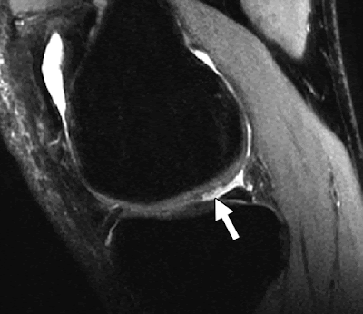
The most common sequences used in clinical practice to evaluate the articular cartilage of the knee joint are 2D fast spin-echo (FSE) sequences with T2-weighted and intermediate-weighted contrast . The advantages of FSE sequences include their high in-plane spatial resolution and their ability to evaluate the menisci, ligaments, and osseous structures in addition to the articular cartilage . Because of their multiple off-resonance pulses, FSE sequences also produce a magnetization transfer effect that can help distinguish between normal articular cartilage and areas of early cartilage degeneration . However, FSE sequences have relatively thick slices and gaps between slices that can obscure small cartilage lesions secondary to partial volume averaging. In addition, T2-weighted FSE sequences have poor contrast between articular cartilage and subchondral bone, which can make it difficult to detect diffuse cartilage thinning and estimate the exact depth of cartilage lesions. Intermediate-weighted FSE sequences are also limited by image blurring secondary to acquisition of high spatial frequencies late in the echo-train and by poor contrast between articular cartilage and synovial fluid . However, image blurring can be reduced by minimizing echo-train length and using short interecho spacing, whereas contrast between articular cartilage and synovial fluid on intermediate-weighted FSE sequences can be improved with the use of fat suppression.
In clinical practice, 3D sequences also have been used to evaluate the articular cartilage of the knee joint. Fat suppression is typically added to these sequences to reduce chemical-shift artifact and to optimize the overall dynamic contrast range of the image. Frequency-selective fat-saturation is the most commonly used method to suppress fat signal . However, 3D sequences with higher cartilage signal-to-noise ratio (SNR) and greater contrast between cartilage and adjacent joint structures can be obtained using recently developed fat-suppression techniques, such as water excitation , linear combination , and iterative decomposition of water and fat with echo asymmetry and least-squares estimates (IDEAL) . By acquiring thin, continuous slices through the knee joint, 3D cartilage imaging sequences can reduce partial volume averaging. The 3D sequences also can be used to create multiplanar reformat images that allow articular cartilage to be evaluated in any orientation after a single acquisition. The disadvantages of 3D cartilage imaging sequences include their long acquisition times; increased susceptibility to artifacts; and limited ability to evaluate the menisci, ligaments, and osseous structures of the knee joint when compared with 2D FSE sequences.
The 3D cartilage imaging sequences can be broadly divided into dark-fluid sequences and bright-fluid sequences on the basis of the signal intensity of synovial fluid. Dark-fluid sequences consist of T1-weighted gradient-recalled echo (GRE) sequences such as spoiled gradient recalled-echo (SPGR) and fast low-angle shot (FLASH). These sequences have been successfully used to evaluate articular cartilage in clinical practice and to perform cartilage volume measurements in osteoarthritis research studies . However, the main disadvantage of using SPGR and FLASH sequences for clinical cartilage imaging is the low signal intensity of synovial fluid, which may decrease the conspicuity of superficial cartilage lesions . Dark-fluid sequences have lower contrast between articular cartilage and synovial fluid than bright-fluid sequences (Kijowski R, et al., presented at the 2008 annual meeting of the International Society of Magnetic Resonance in Medicine). In addition, the surface properties of degenerative cartilage may influence the ability of 3D sequences to detect superficial cartilage lesions. Superficial degeneration shortens the T2 relaxation time of cartilage. For dark-fluid sequences, the T2 shortening of degenerative cartilage has no effect on its signal intensity and contrast relative to synovial fluid. However, for bright-fluid sequences, the effect of T2 shortening is to decrease the signal intensity of degenerative cartilage and thus increase its contrast relative to synovial fluid, which may result in greater conspicuity of superficial cartilage lesions .
Various 3D cartilage imaging sequences with bright synovial fluid have been used to evaluate the articular cartilage of the knee joint. These bright-fluid sequences include dual-echo in the steady-state (DESS); driven equilibrium Fourier transform (DEFT); and T2*-weighted gradient-echo sequences, such as gradient-recalled echo acquired in the steady-state (GRASS) and gradient-recalled echo (GRE). The DESS sequence combines two gradient echoes separated by a refocusing pulse into a single image that increases the signal intensity of both articular cartilage and synovial fluid . The DEFT sequence uses a –90° pulse to return transverse magnetization to the z-axis, which increases the signal intensity of synovial fluid and other tissues with long T1 relaxation times . GRASS and GRE sequences produce images with high-signal-intensity synovial fluid because of coherence of transverse magnetization with secondary T2* weighting . The bright synovial fluid on DESS, DEFT, and T2*-weighted gradient-echo sequences creates an arthrogram-like effect within the knee joint that may increase the conspicuity of superficial cartilage lesions.
Balanced steady-state free precession (SSFP) sequences are additional 3D sequences that have been used to evaluate the articular cartilage of the knee joint. Balanced SSFP sequences include commercially available sequences such as fast imaging employing steady-state acquisition (FIESTA) and true fast imaging with steady-state precession (true FISP) and variants such as fluctuating equilibrium MR (FEMR) [58] and vastly undersampled isotropic-projection steady-state free precession (VIPR-SSFP) [46]. These sequences have high SNR efficiency and produce images of the knee joint with T2-/T1-weighted contrast and bright synovial fluid. Balanced SSFP sequences and their variants have higher cartilage SNR and greater contrast between cartilage and adjacent joint structures than 2D FSE and fat-saturated SPGR sequences [39, 49, 59]. When combined with VIPR radial k-space trajectory and alternating TR fat–water separation, balanced SSFP images of the knee joint with 0.3-mm isotropic resolution can be obtained in as little as 8 minutes (Klaers JK, et al., presented at the 2010 annual meeting of the Society of Magnetic Resonance in Medicine) .
Recently, 3D FSE sequences, such as FSE-Cube (GE Healthcare) and sampling perfection with application oriented contrast using different flip angle evolutions (SPACE, Siemens Healthcare) , have been used to evaluate the articular cartilage of the knee joint. These sequences use variable flip angle modulation to constrain T2 decay over an extended echo-train, which allows intermediate-weighted images of the knee joint with bright synovial fluid to be acquired with minimal blurring. FSE-Cube and SPACE sequences have higher cartilage SNR but lower contrast between cartilage and synovial fluid when compared with 2D FSE sequences . The 3D FSE sequences acquire volumetric data sets with isotropic resolution that allow high-quality multiplanar reformat images to be obtained in any orientation after a single acquisition. However, these sequences have lower in-plane spatial resolution when compared with other 3D cartilage imaging sequences with similar acquisition times, which may reduce the conspicuity of superficial cartilage lesions.


No comments:
Post a Comment